Ultrasound-Guided FNAC/Biopsy
- Home
- Ultrasound-Guided FNAC/Biopsy
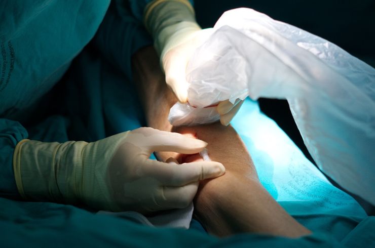
Ultrasound-guided Fine Needle Aspiration Cytology (FNAC) and biopsy are minimally invasive diagnostic procedures used to collect tissue or fluid samples from a specific area in the body. This technique is commonly used to assess lumps, tumors, or other abnormalities in organs such as the thyroid, liver, breast, and lymph nodes.
What Is It?
- FNAC involves using a thin, hollow needle to extract cells from a suspicious mass or organ. The sample is then examined under a microscope to identify any abnormalities or signs of disease.
- Biopsy is similar but may involve a slightly larger needle to obtain a tissue sample. The sample is sent for further histopathological analysis to confirm the presence of any malignant or benign conditions.
Why It Is Done:
- To diagnose cancer or other abnormal growths.
- To evaluate the nature of masses or lumps that can be felt under the skin or identified through imaging.
- To assess conditions affecting internal organs, such as the liver, kidney, or lungs.
How It Works:
- Preparation: The patient is usually positioned comfortably, and an ultrasound machine is used to guide the needle to the precise location of the abnormality.
- Procedure: The doctor numbs the area with a local anesthetic. Using real-time ultrasound imaging, the needle is carefully inserted into the target tissue to extract the sample.
- Post-procedure: Once the sample is collected, it is sent for analysis. Most patients can resume normal activities shortly after the procedure.
Benefits:
- Accuracy: The ultrasound guidance ensures the needle reaches the correct location, reducing the risk of errors.
- Minimal Invasion: The procedure is minimally invasive, causing less discomfort and a faster recovery time compared to traditional surgical biopsies.
- Quick Results: Most results are available within a few days, allowing for quicker diagnosis and treatment.
Risks:
- Although rare, there may be some discomfort, bruising, or bleeding at the site of the biopsy. Infection is also a minimal risk.
Interventions Offered
- Ultrasound-Guided FNAC/Biopsy
- Percutaneous Drainage Procedures
- Soft Tissue & Joint Injections (PRP)
- Spinal Infiltrations
- Nerve Blocks for Pain Management
- Trans Arterial Micro Embolization (TAME)
- PTBD & Biliary Stenting
- PCN & Ureteral Stenting
- Bronchial Artery Embolization for Hemoptysis
- Prostatic Artery Embolization for BPH
- Uterine Artery Embolization for Fibroids
- Varicocele Embolization
- Genicular Artery Embolization for Knee Osteoarthritis
- TACE / TARE for Liver Cancer Treatment
- Thyroid Nodule & Tumor Ablation
- Transjugular Intrahepatic Portosystemic Shunt (TIPS)
- EVLA (Endovenous Laser Ablation) & Sclerotherapy for Varicose Veins
- Thromboaspiration & Catheter-Directed Thrombolysis (CDT) for DVT & Pulmonary Thromboembolism
- Management of Peripheral Artery Disease (PAD)